Gross Anatomy of the Kidney
By the end of this section, you will be able to:
- Describe the external structure of the kidney, including its location, support structures, and covering
- Identify the major internal divisions and structures of the kidney
- Identify the major blood vessels associated with the kidney and trace the path of blood through the kidney
- Compare and contrast the cortical and juxtamedullary nephrons
- Name structures found in the cortex and medulla
- Describe the physiological characteristics of the cortex and medulla
The kidneys lie on either side of the spine in the retroperitoneal space between the parietal peritoneum and the posterior abdominal wall, well protected by muscle, fat, and ribs. They are roughly the size of your fist, and the male kidney is typically a bit larger than the female kidney. The kidneys are well vascularized, receiving about 25 percent of the cardiac output at rest.
 There have never been sufficient kidney donations to provide a kidney to each person needing one. Watch this video to learn about the TED (Technology, Entertainment, Design) Conference held in March 2011. In this video, Dr. Anthony Atala discusses a cutting-edge technique in which a new kidney is “printed.” The successful utilization of this technology is still several years in the future, but imagine a time when you can print a replacement organ or tissue on demand.
External Anatomy
The left kidney is located at about the T12 to L3 vertebrae, whereas the right is lower due to slight displacement by the liver. Upper portions of the kidneys are somewhat protected by the eleventh and twelfth ribs ([link]). Each kidney weighs about 125–175 g in males and 115–155 g in females. They are about 11–14 cm in length, 6 cm wide, and 4 cm thick, and are directly covered by a fibrous capsule composed of dense, irregular connective tissue that helps to hold their shape and protect them. This capsule is covered by a shock-absorbing layer of adipose tissue called the renal fat pad, which in turn is encompassed by a tough renal fascia. The fascia and, to a lesser extent, the overlying peritoneum serve to firmly anchor the kidneys to the posterior abdominal wall in a retroperitoneal position.
.")
On the superior aspect of each kidney is the adrenal gland. The adrenal cortex directly influences renal function through the production of the hormone aldosterone to stimulate sodium reabsorption.
Internal Anatomy
A frontal section through the kidney reveals an outer region called the renal cortex and an inner region called the medulla ([link]). The renal columns are connective tissue extensions that radiate downward from the cortex through the medulla to separate the most characteristic features of the medulla, the renal pyramids and renal papillae. The papillae are bundles of collecting ducts that transport urine made by nephrons to the calyces of the kidney for excretion. The renal columns also serve to divide the kidney into 6–8 lobes and provide a supportive framework for vessels that enter and exit the cortex. The pyramids and renal columns taken together constitute the kidney lobes.
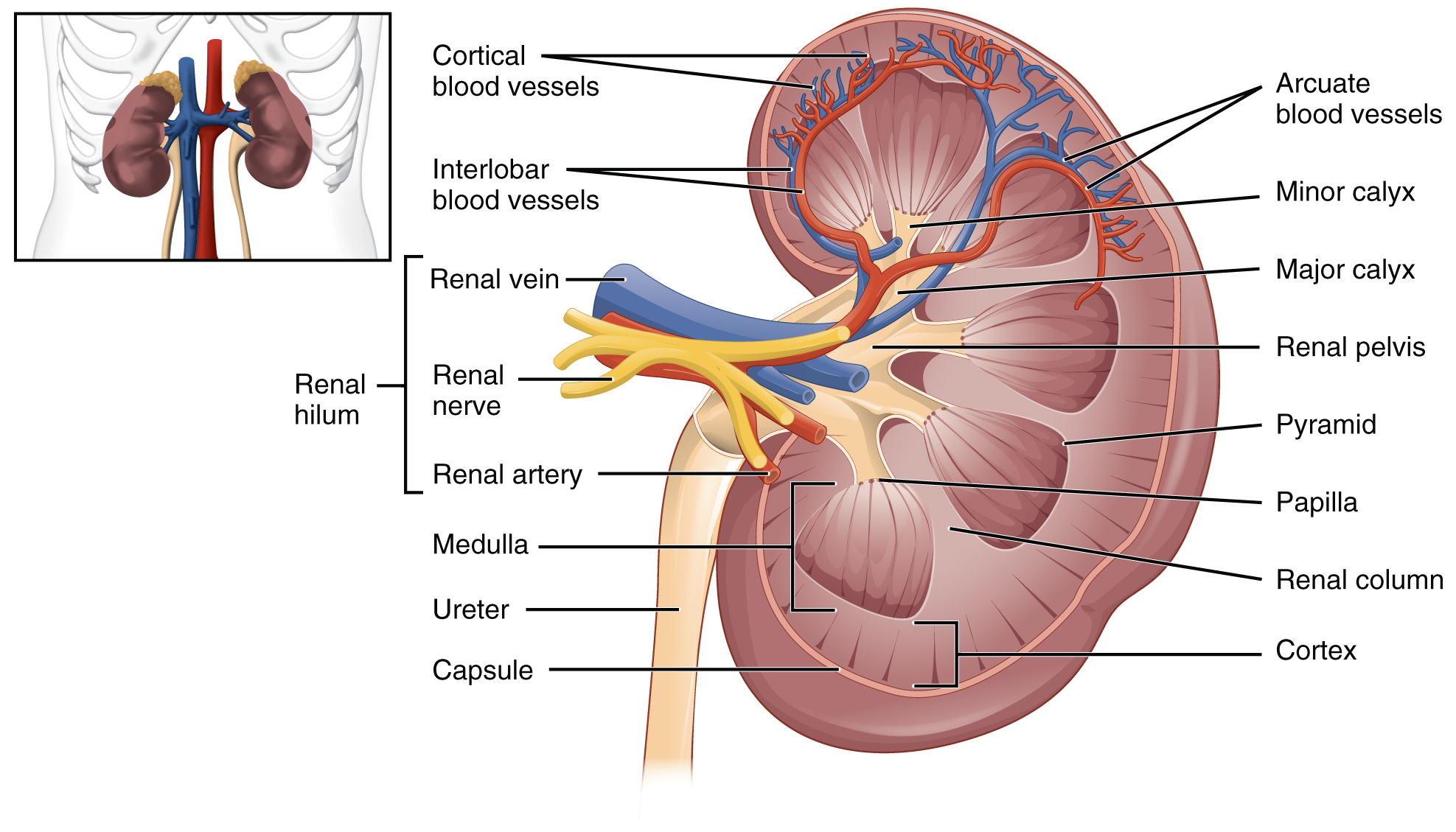
Renal Hilum
The renal hilum is the entry and exit site for structures servicing the kidneys: vessels, nerves, lymphatics, and ureters. The medial-facing hila are tucked into the sweeping convex outline of the cortex. Emerging from the hilum is the renal pelvis, which is formed from the major and minor calyxes in the kidney. The smooth muscle in the renal pelvis funnels urine via peristalsis into the ureter. The renal arteries form directly from the descending aorta, whereas the renal veins return cleansed blood directly to the inferior vena cava. The artery, vein, and renal pelvis are arranged in an anterior-to-posterior order.
Nephrons and Vessels
The renal artery first divides into segmental arteries, followed by further branching to form interlobar arteries that pass through the renal columns to reach the cortex ([link]). The interlobar arteries, in turn, branch into arcuate arteries, cortical radiate arteries, and then into afferent arterioles. The afferent arterioles service about 1.3 million nephrons in each kidney.
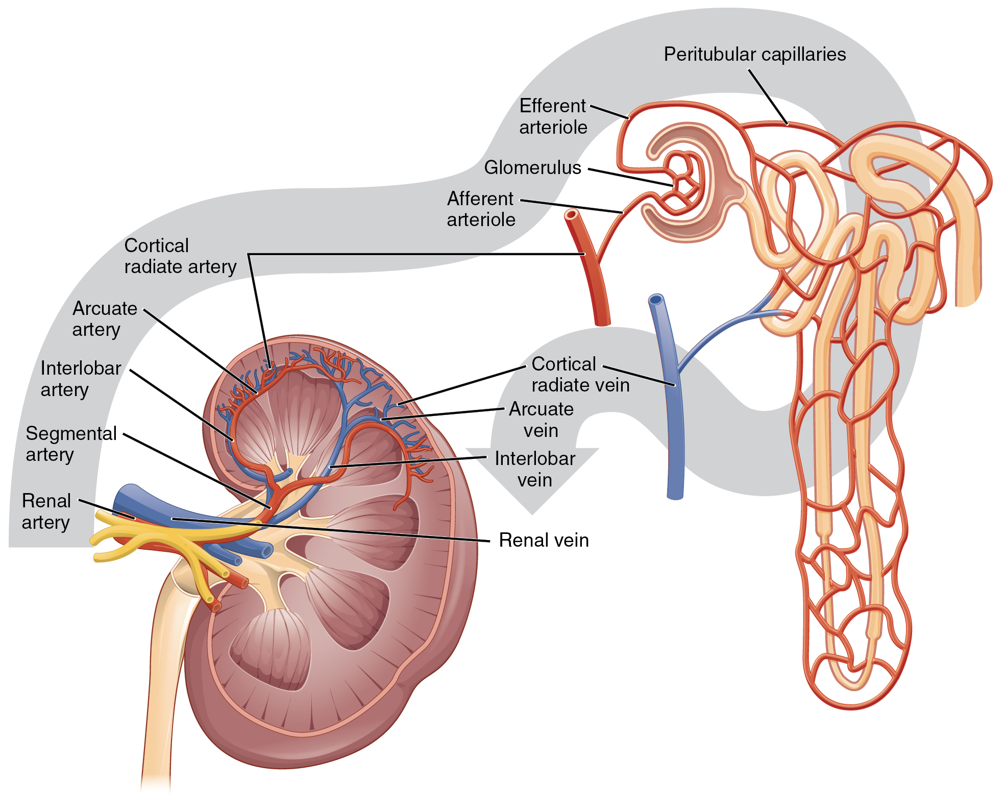
Nephrons are the “functional units” of the kidney; they cleanse the blood and balance the constituents of the circulation. The afferent arterioles form a tuft of high-pressure capillaries about 200 µm in diameter, the glomerulus. The rest of the nephron consists of a continuous sophisticated tubule whose proximal end surrounds the glomerulus in an intimate embrace—this is Bowman’s capsule. The glomerulus and Bowman’s capsule together form the renal corpuscle. As mentioned earlier, these glomerular capillaries filter the blood based on particle size. After passing through the renal corpuscle, the capillaries form a second arteriole, the efferent arteriole ([link]). These will next form a capillary network around the more distal portions of the nephron tubule, the peritubular capillaries and vasa recta, before returning to the venous system. As the glomerular filtrate progresses through the nephron, these capillary networks recover most of the solutes and water, and return them to the circulation. Since a capillary bed (the glomerulus) drains into a vessel that in turn forms a second capillary bed, the definition of a portal system is met. This is the only portal system in which an arteriole is found between the first and second capillary beds. (Portal systems also link the hypothalamus to the anterior pituitary, and the blood vessels of the digestive viscera to the liver.)

 Visit this link to view an interactive tutorial of the flow of blood through the kidney.
Cortex
In a dissected kidney, it is easy to identify the cortex; it appears lighter in color compared to the rest of the kidney. All of the renal corpuscles as well as both the proximal convoluted tubules (PCTs) and distal convoluted tubules are found here. Some nephrons have a short loop of Henle that does not dip beyond the cortex. These nephrons are called cortical nephrons. About 15 percent of nephrons have long loops of Henle that extend deep into the medulla and are called juxtamedullary nephrons.
Chapter Review
As noted previously, the structure of the kidney is divided into two principle regions—the peripheral rim of cortex and the central medulla. The two kidneys receive about 25 percent of cardiac output. They are protected in the retroperitoneal space by the renal fat pad and overlying ribs and muscle. Ureters, blood vessels, lymph vessels, and nerves enter and leave at the renal hilum. The renal arteries arise directly from the aorta, and the renal veins drain directly into the inferior vena cava. Kidney function is derived from the actions of about 1.3 million nephrons per kidney; these are the “functional units.” A capillary bed, the glomerulus, filters blood and the filtrate is captured by Bowman’s capsule. A portal system is formed when the blood flows through a second capillary bed surrounding the proximal and distal convoluted tubules and the loop of Henle. Most water and solutes are recovered by this second capillary bed. This filtrate is processed and finally gathered by collecting ducts that drain into the minor calyces, which merge to form major calyces; the filtrate then proceeds to the renal pelvis and finally the ureters.
Review Questions
The renal pyramids are separated from each other by extensions of the renal cortex called ________.
- renal medulla
- minor calyces
- medullary cortices
- renal columns
The primary structure found within the medulla is the ________.
- loop of Henle
- minor calyces
- portal system
- ureter
The right kidney is slightly lower because ________.
- it is displaced by the liver
- it is displace by the heart
- it is slightly smaller
- it needs protection of the lower ribs
Critical Thinking Questions
What anatomical structures provide protection to the kidney?
Retroperitoneal anchoring, renal fat pads, and ribs provide protection to the kidney.
How does the renal portal system differ from the hypothalamo–hypophyseal and digestive portal systems?
The renal portal system has an artery between the first and second capillary bed. The others have a vein.
Name the structures found in the renal hilum.
The structures found in the renal hilum are arteries, veins, ureters, lymphatics, and nerves.
Glossary
- Bowman’s capsule
- cup-shaped sack lined by a simple squamous epithelium (parietal surface) and specialized cells called podocytes (visceral surface) that participate in the filtration process; receives the filtrate which then passes on to the PCTs
- calyces
- cup-like structures receiving urine from the collecting ducts where it passes on to the renal pelvis and ureter
- cortical nephrons
- nephrons with loops of Henle that do not extend into the renal medulla
- distal convoluted tubules
- portions of the nephron distal to the loop of Henle that receive hyposmotic filtrate from the loop of Henle and empty into collecting ducts
- efferent arteriole
- arteriole carrying blood from the glomerulus to the capillary beds around the convoluted tubules and loop of Henle; portion of the portal system
- glomerulus
- tuft of capillaries surrounded by Bowman’s capsule; filters the blood based on size
- juxtamedullary nephrons
- nephrons adjacent to the border of the cortex and medulla with loops of Henle that extend into the renal medulla
- loop of Henle
- descending and ascending portions between the proximal and distal convoluted tubules; those of cortical nephrons do not extend into the medulla, whereas those of juxtamedullary nephrons do extend into the medulla
- nephrons
- functional units of the kidney that carry out all filtration and modification to produce urine; consist of renal corpuscles, proximal and distal convoluted tubules, and descending and ascending loops of Henle; drain into collecting ducts
- medulla
- inner region of kidney containing the renal pyramids
- peritubular capillaries
- second capillary bed of the renal portal system; surround the proximal and distal convoluted tubules; associated with the vasa recta
- proximal convoluted tubules (PCTs)
- tortuous tubules receiving filtrate from Bowman’s capsule; most active part of the nephron in reabsorption and secretion
- renal columns
- extensions of the renal cortex into the renal medulla; separates the renal pyramids; contains blood vessels and connective tissues
- renal corpuscle
- consists of the glomerulus and Bowman’s capsule
- renal cortex
- outer part of kidney containing all of the nephrons; some nephrons have loops of Henle extending into the medulla
- renal fat pad
- adipose tissue between the renal fascia and the renal capsule that provides protective cushioning to the kidney
- renal hilum
- recessed medial area of the kidney through which the renal artery, renal vein, ureters, lymphatics, and nerves pass
- renal papillae
- medullary area of the renal pyramids where collecting ducts empty urine into the minor calyces
- renal pyramids
- six to eight cone-shaped tissues in the medulla of the kidney containing collecting ducts and the loops of Henle of juxtamedullary nephrons
- vasa recta
- branches of the efferent arterioles that parallel the course of the loops of Henle and are continuous with the peritubular capillaries; with the glomerulus, form a portal system

This work is licensed under a Creative Commons Attribution 4.0 International License.
You can also download for free at http://cnx.org/contents/14fb4ad7-39a1-4eee-ab6e-3ef2482e3e22@11.1
Attribution: